After the chilly trip to Scarborough last week I had just enough time to warm up and don my ‘Civvies’ ready for the trip down to Colchester for the 3 day NERC run ‘Fluorescent Microscopy for Environmental Research course at the University of Essex. I had a basic understanding of what fluorescent microscopy was and how it is used in biology, but my knowledge probably extended to just a few sentences worth of loosely accurate info. I had read papers and seen images of how fluorescent microscopy is used with phytoplankton, and being that all of my data is collated from images I was intrigued to see if I could employ it in my research.
If this course is repeated next Autumn, you really MUST sign up for it! I was blown away by the depth of knowledge of not only the instructors and technicians, but the 15 other Ph.D. students and researchers present. The course itself was presented in a way that covered all bases, from simple questions you may have been afraid to ask (on par with ‘What is a pixel’?) to the very forefront of current bioimaging technology. Following each 2hr lecture was a 2-3hr practical where we got to see first hand how the methods we had just learnt about are used, not to mention play with some shiny (read expensive) equipment from Nikon. I was constantly learning new techniques in the lectures, the lab and at the evening meals where everybody had a particular skillset to bring to the table.
As for fluorescent microscopy applied to my research – well it seems that my swimming organisms are too small and fast to be recorded under fluorescence in a way that would provide me with useful information…although the technology to do this is constantly being updated. Thanks to the expertise of Phillipe Laissiue (http://www.essex.ac.uk/bs/staff/profile.aspx?ID=1205) and Boguslaw Obara (https://www.dur.ac.uk/research/directory/staff/?mode=staff&id=10591), and that of other students, I have come away with exciting ideas on how to optimise my current research and now have a head full of fascinating ideas of how to overcome bioimaging problems in the future.
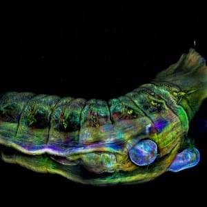
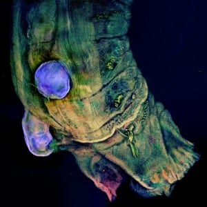
The images below were taken on the course by Bex Summerfield (NHM – London). They display a new species of parasitic copepod (blue sphere) on a polychaete host (from the genus Jasmineira), fixed in 70% ethanol, mounted in vector shield in a glass bottomed petri dish and scanned with Nikon Ti CLSM. The detail is simply mind-blowing!