Teacher one week, pupil the next! Fluorescent Microscopy Course

After the chilly trip to Scarborough last week I had just enough time to warm up and don my ‘Civvies’ ready for the trip down to Colchester for the 3 day NERC run ‘Fluorescent Microscopy for Environmental Research course at the University of Essex. I had a basic understanding of what fluorescent microscopy was and how it is used in biology, but my knowledge probably extended to just a few sentences worth of loosely accurate info. I had read papers and seen images of how fluorescent microscopy is used with phytoplankton, and being that all of my data is collated from images I was intrigued to see if I could employ it in my research.

If this course is repeated next Autumn, you really MUST sign up for it! I was blown away by the depth of knowledge of not only the instructors and technicians, but the 15 other Ph.D. students and researchers present. The course itself was presented in a way that covered all bases, from simple questions you may have been afraid to ask (on par with ‘What is a pixel’?) to the very forefront of current bioimaging technology. Following each 2hr lecture was a 2-3hr practical where we got to see first hand  how the methods we had just learnt about are used, not to mention play with some shiny (read expensive) equipment from Nikon. I was constantly learning new techniques in the lectures, the lab and at the evening meals where everybody had a particular skillset to bring to the table.

As for fluorescent microscopy applied to my research – well it seems that my swimming organisms are too small and fast to be recorded under fluorescence in a way that would provide me with useful information…although the technology to do this is constantly being updated.  Thanks to the expertise of Phillipe Laissiue (http://www.essex.ac.uk/bs/staff/profile.aspx?ID=1205) and Boguslaw Obara (https://www.dur.ac.uk/research/directory/staff/?mode=staff&id=10591), and that of other students, I have come away with exciting ideas on how to optimise my current research and now have a head full of fascinating ideas of how to overcome bioimaging problems in the future.

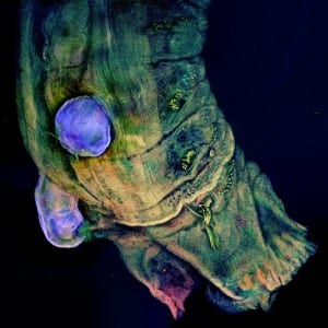
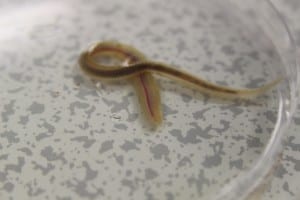
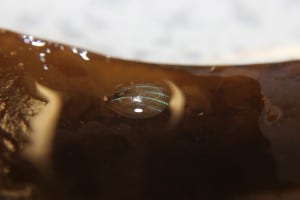
The images below were taken on the course by Bex Summerfield (NHM – London). They display a new species of parasitic copepod (blue sphere) on a polychaete host (from the genus Jasmineira), fixed in 70% ethanol, mounted in vector shield in a glass bottomed petri dish and scanned with Nikon Ti CLSM. The detail is simply mind-blowing!
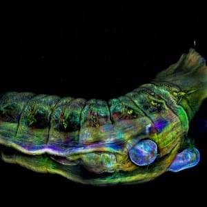
1382893_10154754888280214_1352194327015210488_n 10624580_10154754887890214_2669528823304762238_n


A mostly sunny Scarborough! 1st Year Field course.

The first week in November saw my 4th time demonstrating on the 1st year B.Sc. Marine Biology field trip. Its a week that I always look forward to, where I get to geek out and scour the seashore for the unique and cryptic organisms that inhabit our coastline. Back in 2008 I myself went on the field trip which was (until two years ago) held a further 30mins up the coast at Whitby and Runswick Bay.

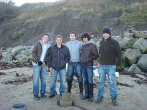
2008 Whitby fieldtrip. Building sandcastles we're not park of the marking criteria (sadly...)

2008 Whitby fieldtrip. Building sandcastles were not part of the marking criteria (sadly…)

Students usually come to the field trip in two groups split 3 days apart, however (somewhat thankfully) we only had one group for 3.5 days. The course is designed to give 1st year students (a month into their degrees) their first taste of marine and coastal ecology, where their days are divided into:

  • Day One – Rocky Shore: An introduction to zonation of species on the rocky shore.
  • Day Two – Sand Shore: Beach profile using theodolite, quadrats and identification of species collected from 30cm sand core.
  • Day Three – Rocky Shore : Profile of the rocky shore using theodolite and identification of species within quadrats.

On the first day, the students are shy and their knowledge of common coastal organisms is limited, as demonstrated with a group quiz that usually yields scores of 1 or 2 out of 10. On the final day the students are given the same quiz, where they often collectively score full marks of both common and scientific names. Its fantastic to see their confidence and social skills develop over just a few days.

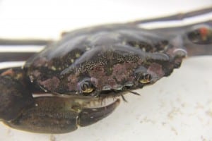
IMG_5536IMG_5562IMG_5566IMG_5572


Teaching students is something I really get a buzz out of, especially when they’re enthusiastic. Here’s hoping I get to continue to do so for many years to come.

Phytoplankton: Natures microscopic marvels

When people ask what I do with my life and I reply that I’m doing my PhD in Marine Biology they immediately expect  that I train dolphins or that I spend 12 hours a day in a wetsuit and jet off to some remote coral reef for 6 months a year. But when I tell them I research microscopic algae I always get the same reaction which usually begins with a disappointed “…Oh”; almost as if all hopes of an exciting and adventurous story about me wrestling a giant squid have been dashed.

At the start of my degree, I too thought of phytoplankton as pointless organisms that merely provided food for other small pointless creatures – and was simply something we all had to learn before being taught about the cool ‘flagship’ species of the marine world…How wrong I was!

I am hopefully going to summarise in a few paragraphs some of the key (but not so well known) facts about phytoplankton that ignited my interest in them, and hopefully will change that disappointed “…Oh” into an “OH!!”.

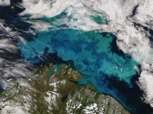
Phytoplankton bloom in the Barents Sea taken from a NASA satellite.

Phytoplankton bloom in the Barents Sea taken from a NASA satellite.

Thank Phytoplankton for every other breath you take!

The average human breathes 12 times a minute, and in the 60 seconds it took for you to read the above text – the phytoplankton themselves theoretically provided you with oxygen for roughly 30 of those seconds. Field et al. (1998) calculated that phytoplankton produce 50% of the worlds’ oxygen supply; however other researchers have suggested that this value could be as high as 70%.

So are phytoplankton really that important to us? The answer is clearly Yes!

Not just a green speck.

Think phytoplankton all look the same? Think again! Size varies tremendously between species – over 4 orders of magnitude! This scale difference between the smallest (Prochlorococcus) and largest (Trichodesmium) genus would be similar to the scale difference between a Red Ant and a Blue Whale.

Picture an obscure shape in your minds eye; chances are a species of phytoplankton exists in this format. The array of morphological configurations of these microscopic plants is truly astounding and often act to optimise a cells position within the water column. Some species have opted for more simple ellipsoid shapes; but coupled with their own methods of propulsion some can swim a distance of 30 body lengths in a single second!

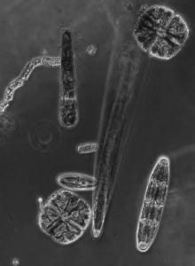
freshwater diatoms and desmids

A small selection of freshwater diatoms and desmids.

Creators of Cliffs and Clouds

The White Cliffs of Dover, created by a plant…a plant that could pass through the eye of a needle with ease…doesn’t sound possible does it! But it’s true! Coccolithophores are a genus of phytoplankton that have characteristic calcium carbonate plates encompassing their surface. Over eons, these phytoplankter sank deep down into the sediment before being forced upwards to form the famous cliffs.

Although dependent on the sun, phytoplankton are sensitive to strong UV rays. When exposed to intense sunlight the cells create their own sunblock from DMSP. Bacteria then break this chemical down into DMS which filters from the ocean into the air, where it breaks down again to form tiny dust-like particles. These tiny particles are just the right size for water to condense on, which is the beginning of how clouds are formed.


So next time you see a murky green pond, remember just how much these ancient plants have impacted the world we live in today.